Institute of Atomic and Molecular Sciences
The Institute conducts research at the atomic and molecular scales, advancing fundamental understanding of physical, chemical, and biological phenomena through the integration of theory and experiment.
Academic Honors

NSTC SEED–Young Scholars Program
Congratulations to Dr. Liang-Yan Hsu on receiving the 2026 NSTC SEED–Young Scholars Program.
2026/07/01

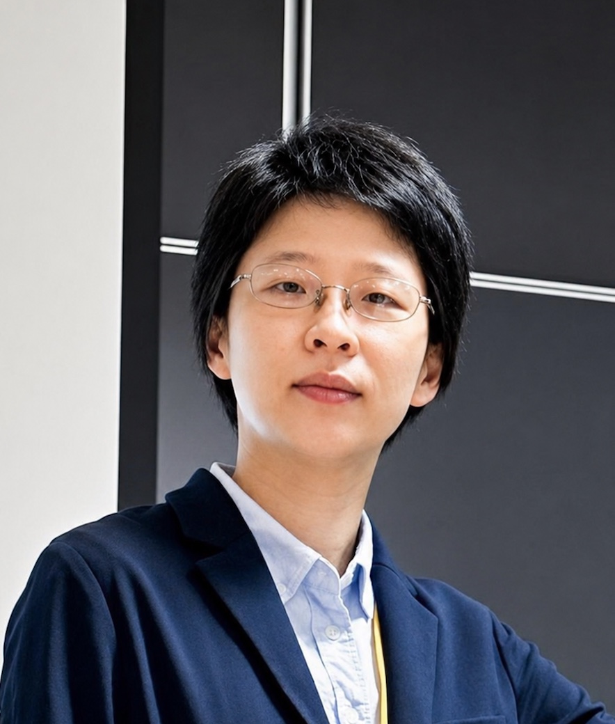
Leap Fellowship of FAOS
Congratulations to Dr. Charles Pin-Kuang Lai on receiving the 2026 Leap Fellowship from the Foundation for the Advancement of Outstanding Scholarship.
2026/02/12
2025 APS Fellow
Congratulations to our jointly appointed research fellow, Distinguished Professor Minn-Tsong Lin, on being elected a 2025 Fellow of the American Physical Society.
2025/10/16
NSTC Outstanding Research Award
Congratulations to Dr. Liang-Yan Hsu on receiving the 2024 Outstanding Research Award from the National Science and Technology Council.
2025/02/26
NSTC Dr. Ta-You Wu Memorial Award
Congratulations to Dr. Pei-Ling Luo on receiving the 2024 Dr. Ta-You Wu Memorial Award from the National Science and Technology Council.
2024/08/28

TTL Biomedical Foundation Young Scholar Award
Congratulations to Dr. Chia-Lung Hsieh on receiving the 20th Young Scholar Award in Biomedical Technology from the Tien-Te Lee Biomedical Foundation.
2024/07/23

NSTC Oustanding Research Award
Congratulations to Dr. Charles Pin-Kuang Lai on receiving the 2023 Outstanding Research Award from the National Science and Technology Council.
2024/02/27

NSTC Outstanding Research Award
Congratulations to Dr. Jer-Lai Kuo receiving the 2023 Outstanding Research Award from the National Science and Technology Council.
2024/2/27