Correlative Light-Electron Microscopy of Lipid-Encapsulated Fluorescent Nanodiamonds for Nanometric Localization of Cell Surface Antigens
Feng-Jen Hsieh,1-3,§ Yen-Wei Chen,1,§ Yao-Kuan Huang,4 Hsien-Ming Lee,5 Chun-Hung Lin,2,3,6 and Huan-Cheng Chang*,1,7
1Institute of Atomic and Molecular Sciences, Academia Sinica, Taipei 106, Taiwan
2Taiwan International Graduate Program – Chemical Biology and Molecular Biophysics, Academia Sinica, Taipei 115, Taiwan
3Department of Biochemical Sciences, National Taiwan University, Taipei 106, Taiwan
4Institute of Cellular and Organismic Biology, Academia Sinica, Taipei 115, Taiwan
5Institute of Chemistry, Academia Sinica, Taipei 115, Taiwan
6Institute of Biological Chemistry, Academia Sinica, Taipei 115, Taiwan
7Department of Chemical Engineering, National Taiwan University of Science and Technology, Taipei 106, Taiwan
§These two authors contribute equally to this work.
Anal. Chem. 90, 1566–1571 (2018).
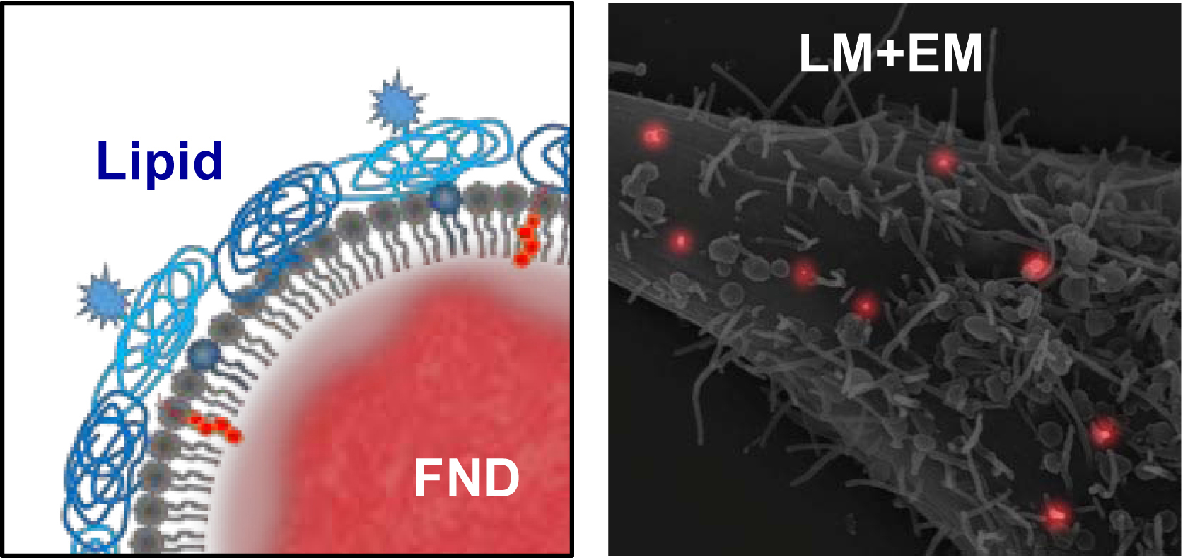
Containing an ensemble of nitrogen-vacancy centers in crystal matrices, fluorescent nanodiamonds (FNDs) are a new type of photostable markers that have found wide applications in light microscopy. The nanomaterial also has a dense carbon core, making it visible to electron microscopy. Here, we show that FNDs encapsulated in biotinylated lipids (bLs) are useful for sub-diffraction imaging of antigens on cell surface with correlative light-electron microscopy (CLEM). The lipid encapsulation enables not only good dispersion of the particles in biological buffers but also high specific labeling of live cells. By employing the bL-encapsulated FNDs to target CD44 on HeLa cell surface through biotin-mediated immunostaining, we obtained the spatial distribution of these antigens by CLEM with a localization accuracy of ~50 nm in routine operations. A comparative study with dual-color imaging, in which CD44 was labeled with FND and MICA/MICB was labeled with Alexa Fluor 488, demonstrated the superior performance of FNDs as fluorescent fiducial markers for CLEM of cell surface antigens.
 Institute of Atomic and Molecular Sciences Academia Sinica

