Fluorescent Nanodiamonds Enable Quantitative Tracking of Human Mesenchymal Stem Cells in Miniature Pigs
Long-Jyun Su,1,2,† Meng-Shiue Wu,3,† Yuen Yung Hui,1,† Be-Ming Chang,1 Lei Pan,1 Pei-Chen Hsu,4 Yit-Tsong Chen,2 Hong-Nerng Ho,5 Yen-Hua Huang,6 Thai-Yen Ling,*,3 Hsao-Hsun Hsu,*,4 and Huan-Cheng Chang*,1,7
1Institute of Atomic and Molecular Sciences, Academia Sinica, Taipei 106, Taiwan
2Department of Chemistry, National Taiwan University, Taipei 106, Taiwan
3Department of Pharmacology, National Taiwan University, Taipei 100, Taiwan
4Department of Surgery, College of Medicine and the Hospital, National Taiwan University, Taipei 100, Taiwan
5Department of Obstetrics and Gynecology, College of Medicine and the Hospital, National Taiwan University, Taipei 100, Taiwan
6Department of Biochemistry and Molecular Cell Biology, Graduate Institute of Medical Sciences, Centre for Cell Therapy and Regeneration Medicine, and International PhD Program for Cell Therapy and Regeneration Medicine, Taipei Medical University, Taipei 110, Taiwan
7Department of Chemical Engineering, National Taiwan University of Science and Technology, Taipei 106, Taiwan
†These authors contributed equally to this work.
Sci. Rep. 7, 45607 (2017) (11pp).
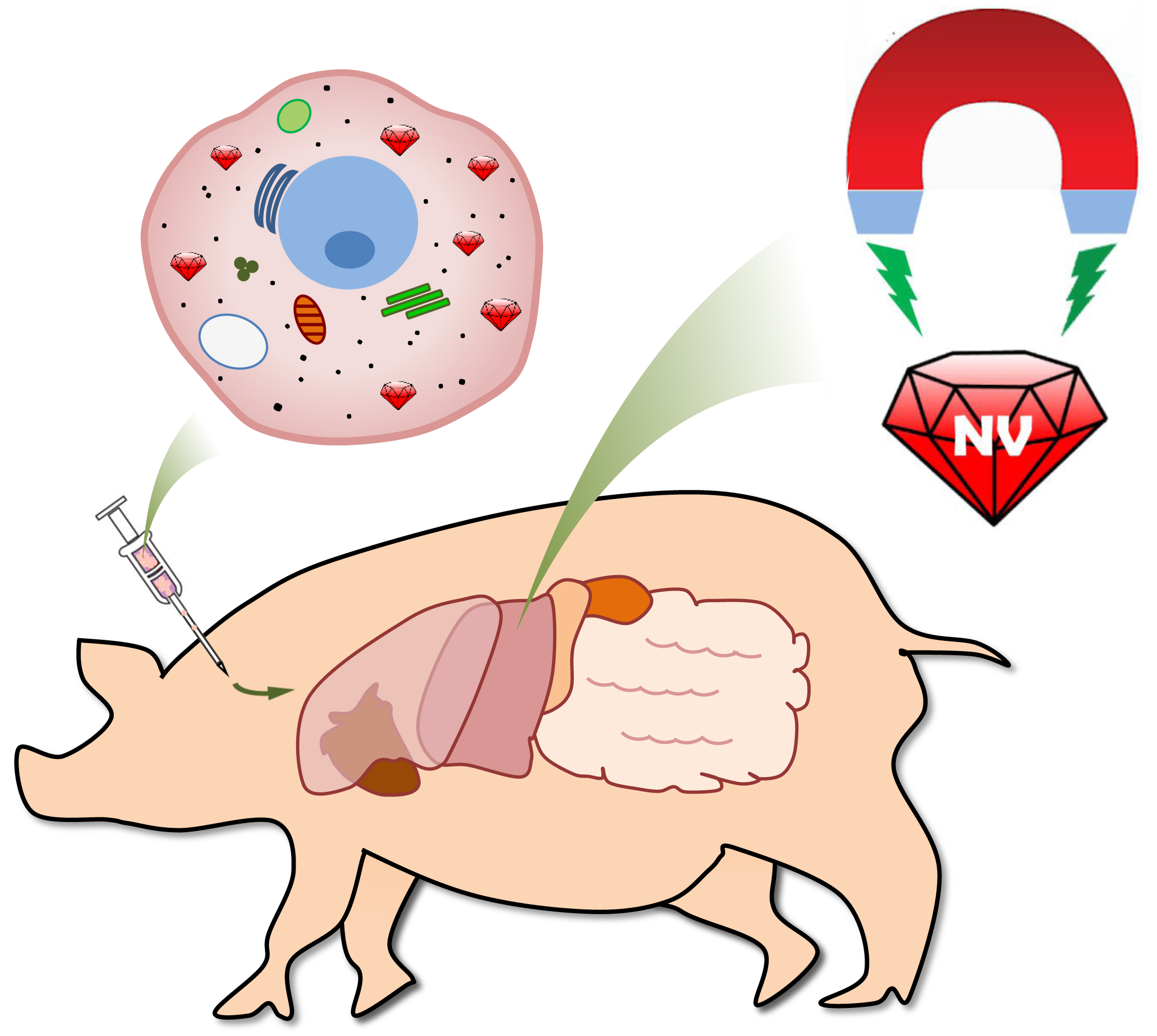
Cell therapy is a promising strategy for the treatment of human diseases. While the first use of cells for therapeutic purposes can be traced to the 19th century, there has been a lack of general and reliable methods to study the biodistribution and associated pharmacokinetics of transplanted cells in various animal models for preclinical evaluation. Here, we present a new platform using albumin-conjugated fluorescent nanodiamonds (FNDs) as biocompatible and photostable labels for quantitative tracking of human placenta choriodecidual membrane-derived mesenchymal stem cells (pcMSCs) in miniature pigs by magnetic modulation. With this background-free detection technique and time-gated fluorescence imaging, we have been able to precisely determine the numbers as well as positions of the transplanted FND-labeled pcMSCs in organs and tissues of the miniature pigs after intravenous administration. The method is applicable to single-cell imaging and quantitative tracking of human stem/progenitor cells in rodents and other animal models as well.
 Institute of Atomic and Molecular Sciences Academia Sinica

