Fluorescent Nanodiamond: A Versatile Tool for Long-Term Cell Tracking, Super-Resolution Imaging, and Nanoscale Temperature Sensing
Wesley Wei-Wen Hsiao,1 Yuen Yung Hui,1 Pei-Chang Tsai,1 and Huan-Cheng Chang*,1,2
1Institute of Atomic and Molecular Sciences, Academia Sinica, Taipei 106, Taiwan
2Department of Chemical Engineering, National Taiwan University of Science and Technology, Taipei 106, Taiwan
Acc. Chem. Res. 49, 400–407 (2016).
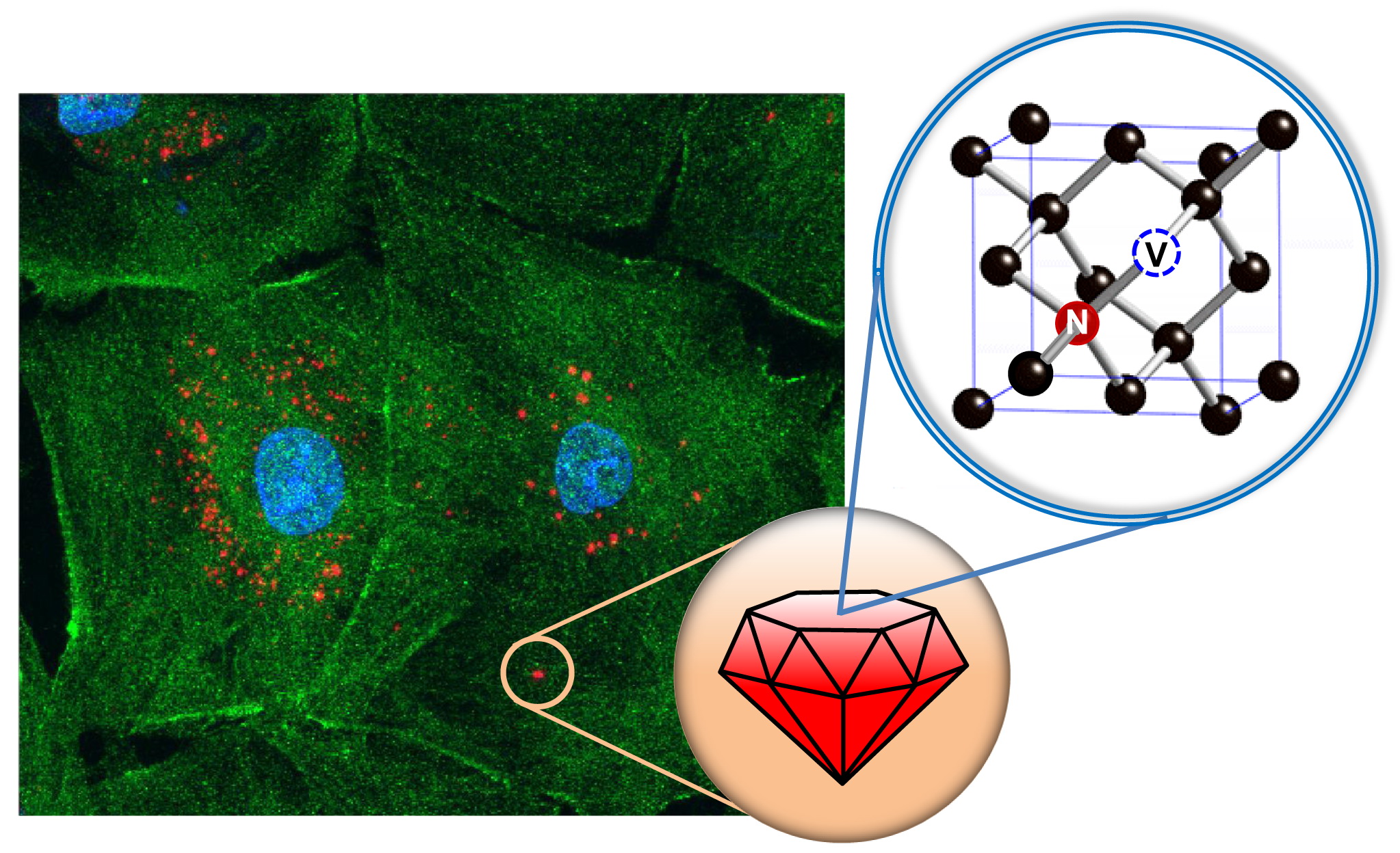
Fluorescent nanodiamond (FND) has recently played a central role in fueling new discoveries in interdisciplinary fields spanning biology, chemistry, physics, and materials sciences. The nanoparticle is unique in that it contains a high density ensemble of negatively charged nitrogen-vacancy (NV–) centers with outstanding optical and magnetic properties. Firstly, NV– has an absorption maximum at ~550 nm and when exposed to green-orange light, it emits bright fluorescence at ~700 nm with a lifetime of longer than 10 ns. These spectroscopic properties are little affected by surface modification and allow background-free imaging of FNDs in tissue sections. Next, as an artificial atom in the solid state, the NV– center is perfectly photostable, without photobleaching and blinking. Therefore, the NV-containing FND is suitable as a contrast agent for super-resolution imaging by stimulated emission depletion (STED). Last, the NV– center in diamond is an atom-like quantum system with a total electron spin of 1 and the ground states of the spins show a crystal field splitting of 2.87 GHz, separating the ms = 0 and ±1 sublevels. Nanothermometry with both high spatial and temporal resolution can be achieved with a technique known as optically detected magnetic resonance (ODMR). This account provides a summary of the recent advances in FND-enabled technologies with a special focus on long-term cell tracking, super-resolution imaging, and nanoscale temperature sensing. These emerging and multifaceted technologies are in synchronicity with modern imaging modalities.
 Institute of Atomic and Molecular Sciences Academia Sinica

